41 compact bone diagram labeled
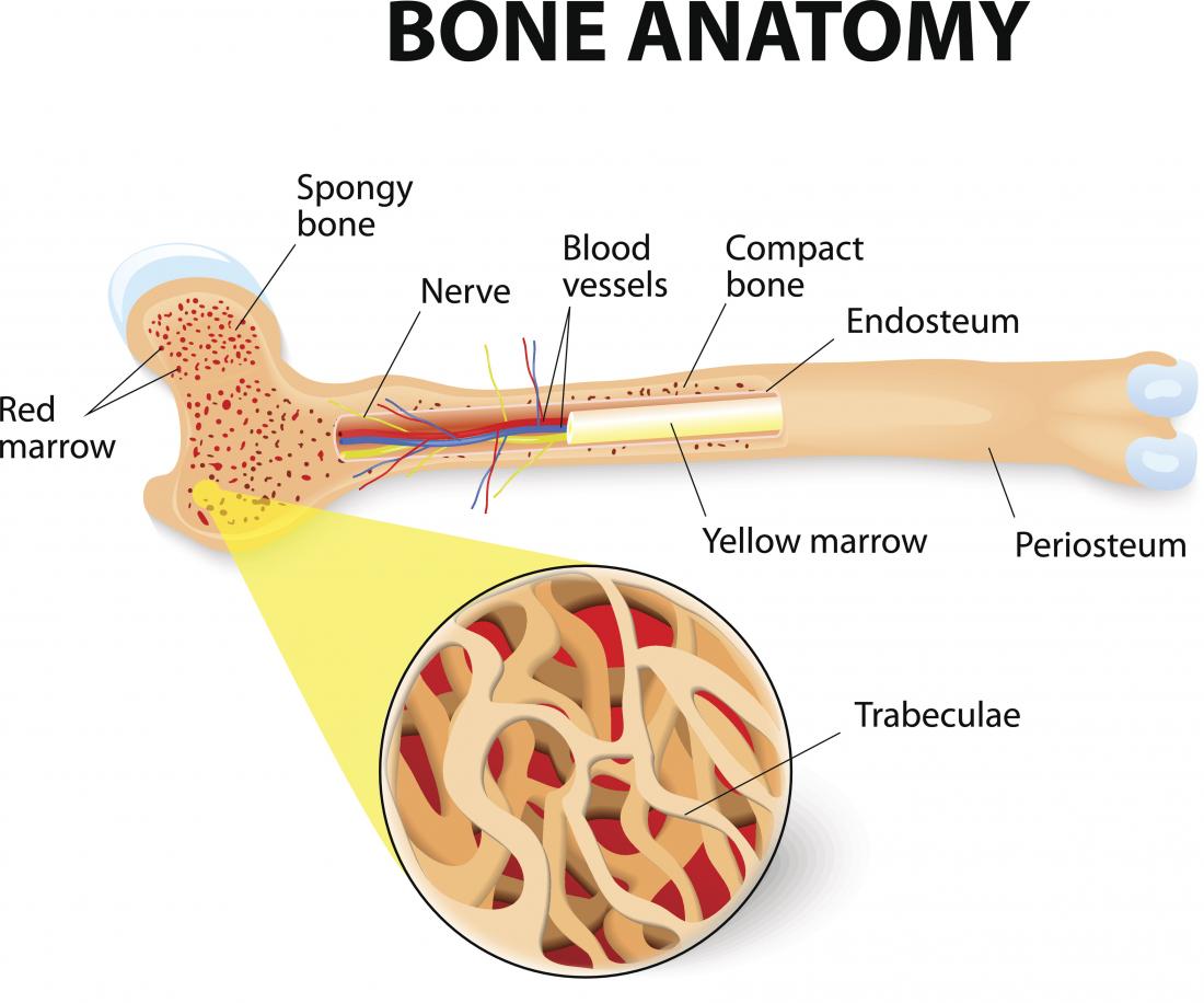
Femur bone anatomy made easy using a labeled diagram of the main parts of the thigh bone along with their location. Includes anatomy of the femur quiz. Fractures to the femur and hip bone can occur and knowing the anatomy will help with management. Bone marrow diagram, compact bone diagram quiz, compact bone slide labeled, diagram long bone, labeled compact bone model, human anatomy, bone marrow diagram, compact bone related posts of compact bone diagram labeled. Illustration about compact bone, also called cortical bone, is the hard, stiff, smooth, thin, white bone tissue that surrounds ...
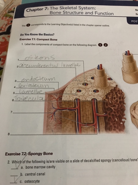
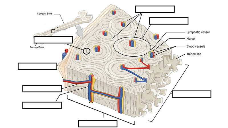
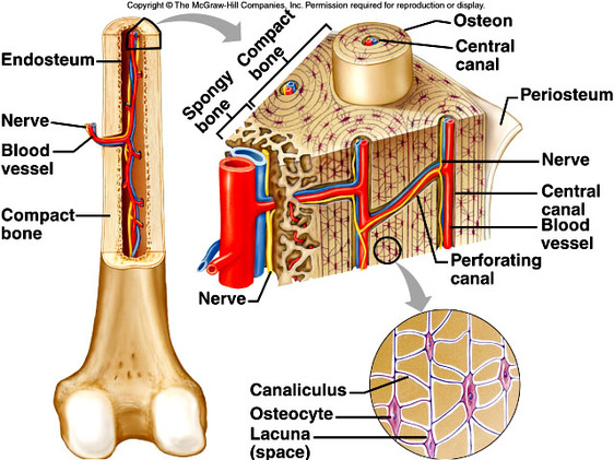
Anatomy Compact bone diagram. STUDY. Flashcards. Learn. Write. Spell. Test. PLAY. Match. Gravity. Created by. mmacfarlane20. Terms in this set (8) spongy bone (contains red marrow) compact bone (has osteons) osteon. osteonic canal. contains vessels and nerves. periosteum. outer covering of bone. perforating canal.
Compact bone diagram labeled
22.08.2021 · In the labeled diagram, I showed you the skull bone, vertebrae (cervical to caudal), ribs, sternum, wing bones, and leg bones from a chicken. You may also get help from the video that I will add at the end of this article. That video might help you to identify all the bones of a chicken. Chicken skeleton anatomy diagram. Chicken bone anatomy. In each bone from chicken skeleton anatomy, I will ... 28.01.2021 · The cranium is the part of the skull that protects the brain, and it is made up of eight bones, and it also supports the structure of the face. Our cranial bones do not fuse but remain distinct and separate throughout our lives. See if you can identify the different cranial bones that are labeled in the cranium bone quiz below. A diagram of the anatomy of a bone, showing the compact bone. Compact and cancellous — or spongy — bone are the two types of tissue found within most bones. Due to its function, compact bone is also referred to as strong bone; due to its structure, it is referred to as cortical bone. The two tissues serve different purposes in bones, with ...
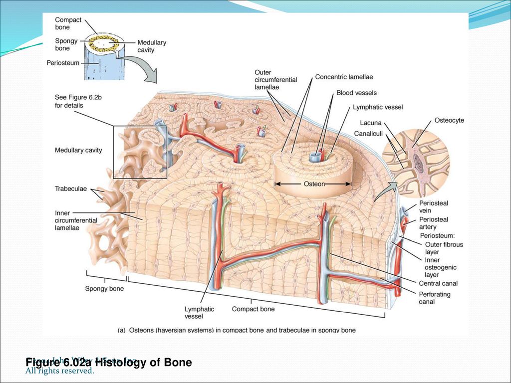
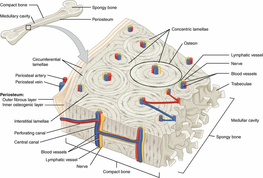
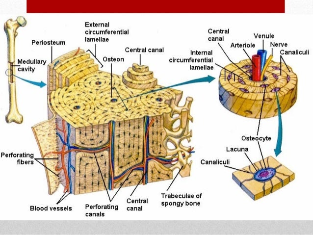
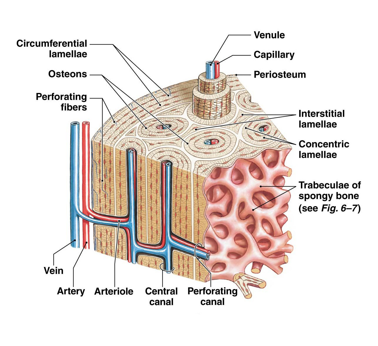
Compact bone diagram labeled. In long bones, as you move from the outer cortical compact bone to the inner medullary cavity, the bone transitions to spongy bone. Figure 6.3.6 - Diagram of Compact Bone: (a) This cross-sectional view of compact bone shows several osteons, the basic structural unit of compact bone. Compact Bone Definition. Compact bone, also known as cortical bone, is a denser material used to create much of the hard structure of the skeleton.As seen in the image below, compact bone forms the cortex, or hard outer shell of most bones in the body.The remainder of the bone is formed by cancellous or spongy bone.. Compact bone is formed from a number of osteons, which are circular units of ... bone. Note osteoclastic bone resorption (resorption phase) at the lower left (solid arrows) and new bone formation (formation phase) at the upper right (open arrows). Undecalcified, 5-µm toluidine blue stained. Slide 12. Same bone section (Slide 11) at higher magnification. SkeleTech - Bone Histology Slide Set Page 4 of 7 2001 each bone is covered with a thin connective tissue membrane called the periosteum, which contains numerous blood vessels, nerves, and lymphatic vessels. The dense and hard exterior surface bone is called cortical or compact bone . Cancellous or spongy bone is found inside the bone. As its name indicates, spongy bone has spaces in
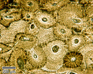
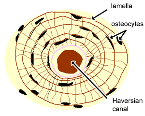
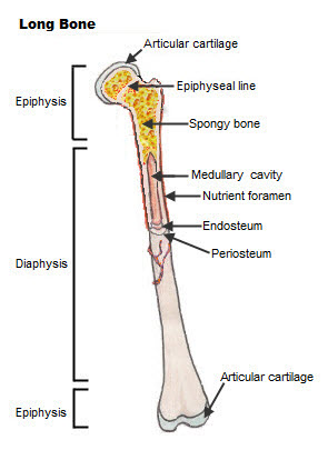
Compact bone makes up the wall of the diaphysis; the epiphyses are filled with spongy bone to reduce the weight of the skeleton. The human body is full of many tiny bones, but there are only 18-20 bones that play a major part in the structure of our skeleton. Worksheets are very critical for every student to practice his her concepts. e. This packet is still . Free multiple-choice quizzes on ... Bodytomy provides a labeled diagram of the Haversian system to help you The terms 'Haversian system' or 'osteon' refer to the basic. osteon diagram TS. The diagram above shows a transverse view of an osteon ( Haversian system) - the basic unit of compact bone. diagram of haversian canal. This video was produced to help students of human anatomy ... 29.07.2020 · Compact bone is made of a matrix of hard mineral salts reinforced with tough collagen fibers. Many tiny cells called osteocytes live in small spaces in the matrix and help to maintain the strength and integrity of the compact bone. Deep to the compact bone layer is a region of spongy bone where the bone tissue grows in thin columns called trabeculae with spaces for red bone marrow in … Compact bone histology slide structure with diagram. Do you want to learn the details of the histology of compact bone with labelled diagram and authentic slide images? Good, here in this part, I am going to describe the structure of compact bone. In compact bone, you will find the three bone lamellar system in an orderly manner - #1.
Start studying Compact Bone Labeling. Learn vocabulary, terms, and more with flashcards, games, and other study tools. A diagram of the anatomy of a bone, showing the compact bone. Human bone generally comprises osseous tissue, an outer coating called a periosteum, and bone marrow. The two main structural components typically include spongy bone on the interior, with an outer layer of compact bone. Usually found in long bones of the body, it consists of units ... Honors Anatomy Chapter 6. 58 terms. Lucia_Macias. Compact bone. 9 terms. Lucia_Macias. Diagram of Bone. 4 terms. Lucia_Macias. Lab ex. 17 part 2. 25 terms. BruceyGirl12. OTHER SETS BY THIS CREATOR. Pharm II Final Exam. 72 terms. ... Start studying Compact Bone Under Microscope. Learn vocabulary, terms, and more with flashcards, games, and other ... Compact bone is the denser, stronger of the two types of bone tissue ( [link] ). It can be found under the periosteum and in the diaphyses of long bones, where it provides support and protection. Diagram of Compact Bone. (a) This cross-sectional view of compact bone shows the basic structural unit, the osteon.
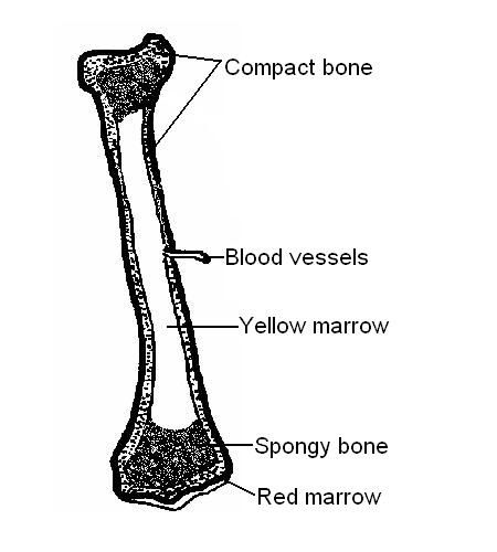
(On Textbook Page Diagrams Note Only Highlighted Labels) Compact Bone. Osteon Model Lacunae Canaliculi Osteocyte. Concentric Lamellae Interstitial Lamellae Central Canal Lacuna Osteocyte Canaliculus. Anatomy of a Long Bone Proximal Epiphysis Diaphysis Distal Epiphysis Compact Bone Spongy Bone Medullary Cavity. Cervical Vertebrae: Atlas ...
Compact bone is the denser, stronger of the two types of bone tissue ( (Figure) ). It can be found under the periosteum and in the diaphyses of long bones, where it provides support and protection. Diagram of Compact Bone. (a) This cross-sectional view of compact bone shows the basic structural unit, the osteon.
05.10.2021 · In endochondral ossification, bone is formed by replacing the calcified cartilage.In this article, I will discuss the detailed process of endochondral ossification with labeled diagrams. Hi there, welcome back again, and many, many thanks for getting into this article. I hope this article is going to be one of the best and easiest articles on the internet to learn the whole endochondral ...
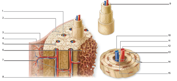
Which of the labeled structures in the diagram are fragments of older osteons that have been partially destroyed during bone rebuilding or growth? A- Interstitial llamanae. ... bone remodeling occurs and compact bone replaces the spongy bone around the periphery of the fracture site.
Label parts of compact bone Learn with flashcards, games, and more — for free.
This is an online quiz called Structure of Compact Bone. There is a printable worksheet available for download here so you can take the quiz with pen and paper. From the quiz author. Structure of Compact Bone Your Skills & Rank. Total Points. 0. Get started! Today's Rank--0. Today 's Points.
05.04.2017 · The image shows a diagram of a human endoskeleton with the major bones labeled. Fish within the class chondrichthyes (sharks, rays and chimaeras) have an endoskeleton; although, rather than bone, their skeletons are made up of cartilage, muscle and connective tissues. While the majority of invertebrates have a non-cartilaginous exoskeleton, a select few invertebrates have endoskeletons ...
05.04.2021 · Depending on the size and distribution of the spaces in the matrix, regions of the bone can be either compact or spongy. Compact bone tissue contains few spaces and is the strongest form of bone tissue. Spongy bone tissue has numerous spaces with some spaces filled with red bone marrow. C. Blood. Created with BioRender.com. Blood is a specialized fluid connective tissue consisting of …
Module 6.3: Long bones transmit forces along the shaft and have a rich blood supply Long bone features Epiphysis (expanded area at each end of the bone) •Consists largely of spongy bone (trabecular bone) •Outer covering of compact bone (cortical bone) -Strong, organized bone •Articular cartilage -Covers portions of epiphysis that form
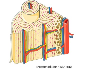
Given below is a labeled diagram to help you understand the structure of compact long bones, as well as the microscopic structure or histology of the Haversian system of compact bones.Structure of a Long Bone Only compact bones have osteons as a basic structural unit; spongy bones don't have osteons.
In the last article, I described the compact bone histology with labeled diagram and real slide pictures. This short article will describe the spongy bone histology and labeled diagram and real slide pictures. Hey there, welcome back again to anatomy learner and thank you so much for getting into this article.
Compact bone, also called cortical bone, is the hard, stiff, smooth, thin, white bone tissue that surrounds all bones in the human body. It is also called osseous tissue or cortical bone and it provides structure and support for an organism as part of its skeleton, in addition to being a location for the storage of minerals like calcium.About 80% of the weight of the human skeleton comes from ...
compact bone, also called cortical bone, dense bone in which the bony matrix is solidly filled with organic ground substance and inorganic salts, leaving only tiny spaces (lacunae) that contain the osteocytes, or bone cells.Compact bone makes up 80 percent of the human skeleton; the remainder is cancellous bone, which has a spongelike appearance with numerous large spaces and is found in the ...
Structure of Bone Tissue. There are two types of bone tissue: compact and spongy.The names imply that the two types differ in density, or how tightly the tissue is packed together. There are three types of cells that contribute to bone homeostasis.Osteoblasts are bone-forming cell, osteoclasts resorb or break down bone, and osteocytes are mature bone cells.
You’ve just entered the laboratory of the mad scientist, Dr. Build-A-Bone! Dr. Build-A-Bone has dedicated his life to discovering what mysterious substances are in bones, and to developing a process for growing new bone. For years scientists have been searching for his laboratory — now you are the lucky one who has found it! But you don’t ...
Compact bone and spongy/cancellous bone are the two types of bones in the human body. The former makes up about 80% of the bones of the human body, while the latter constitutes the remaining 20%. The compact bones form the hard exterior of the bones, whereas the spongy bones have several pores that are filled with nerves and blood vessels. The terms ‘Haversian system’ or ‘osteon’ refer ...
Compact bone accounts for 80% of the bones in the human body. Compact bone, as opposed to spongy bone, is made of cylindrical units, called osteons, that are tightly formed together. As compact ...
In anatomy, the scapula (plural scapulae or scapulas), also known as the shoulder bone, shoulder blade, wing bone, speal bone or blade bone, is the bone that connects the humerus (upper arm bone) with the clavicle (collar bone). Like their connected bones, the scapulae are paired, with each scapula on either side of the body being roughly a mirror image of the other.
The diagram above shows a transverse view of an osteon (Haversian system) - the basic unit of compact bone. The diagram above shows a longitudinal view of an osteon. Some, mostly older, compact bone is remodelled to form these Haversian systems (or osteons).
Anatomy of a Bone -Coloring . EPIPHYSIS (a) - at the ends of the bone (do not color) The epiphysis has a thin layer of compact bone, while internally the bone is cancellous. The epiphysis is capped with articular cartilage. EPIPHYSEAL LINE (j) - purple The epiphyseal line or disk is also called the growth plate, it is found on both ends of the ...
A diagram of the anatomy of a bone, showing the compact bone. Compact and cancellous — or spongy — bone are the two types of tissue found within most bones. Due to its function, compact bone is also referred to as strong bone; due to its structure, it is referred to as cortical bone. The two tissues serve different purposes in bones, with ...
28.01.2021 · The cranium is the part of the skull that protects the brain, and it is made up of eight bones, and it also supports the structure of the face. Our cranial bones do not fuse but remain distinct and separate throughout our lives. See if you can identify the different cranial bones that are labeled in the cranium bone quiz below.
22.08.2021 · In the labeled diagram, I showed you the skull bone, vertebrae (cervical to caudal), ribs, sternum, wing bones, and leg bones from a chicken. You may also get help from the video that I will add at the end of this article. That video might help you to identify all the bones of a chicken. Chicken skeleton anatomy diagram. Chicken bone anatomy. In each bone from chicken skeleton anatomy, I will ...














0 Response to "41 compact bone diagram labeled"
Post a Comment