41 wiggers diagram aortic stenosis
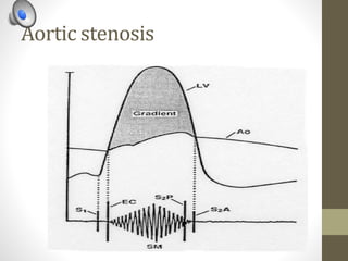
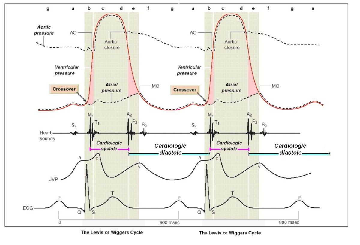
The Cardiac Cycle - University of Cape Town The “Wiggers diagram” Prof. Justiaan Swanevelder Dept of Anaesthesia & Perioperative Medicine University of Cape Town Each cardiac cycle consists of a period of relaxation (diastole) followed by ventricular contraction (systole). During diastole the ventricles are relaxed to allow filling. In systole the right and left CV Physiology | Valvular Stenosis Stenosis of either atrioventricular valves (tricuspid, mitral) or outflow tract valves (pulmonic, aortic) leads to an elevated pressure gradient across the valve as blood is flowing through the valve opening. This increased pressure gradient is expressed as an increase in the pressure proximal to the valve and a small fall in pressure distal to the valve.
The Wright table of the cardiac cycle: a stand-alone ... The Wright table is introduced as a novel tool for teaching and learning the cardiac cycle. It supplements the nearly 100-yr-old Wiggers diagram, which is information rich but difficult for many students to learn. The Wright table offers a compact presentation of information, viewable both in terms …

Wiggers diagram aortic stenosis
Wiggers Diagram Tutorial - XpCourse Wiggers diagram A Wiggers diagram is a standard diagram used in cardiac physiology named after Dr. Carl J. Wiggers. The X axis is used to plot time, while the Y axis contains all of the following on a single grid: More Courses ›› View Course (PPT) Ciclo cardiaco y diagrama de wiggers | Gabriela ... Now PDF Aortic stenosis s2 Aortic stenosis s2 Soft and single s2 aortic stenosis. Paradoxical split s2 aortic stenosis. ... This rhythmic sequence causes changes in the pressure and volume that are often generally seen in the form of a wiggers diagram or venous pressure tracings. Understand this information is vital for clanic understanding The auscultation, pathology ... PDF PHONOCARDIOGRAM OFAORTIC STENOSIS - Heart Thephonocardiogramofaortic stenosis showsasystolic murmurofcharacteristic shapebothin the aortic andmitral areas. Themurmuris smallat first, rising to apeakin mid-systoleandthen decreasing so that it is small or absent before reaching the second sound. In mitral valve disease the systolic[murmur tends to fill systole more evenly or to be mainly ...
Wiggers diagram aortic stenosis. Wiggers Diagram Aortic Regurgitation - schematron.org May 10, 2018 · Diagram of the ascending and descending aorta illustrating . Stewart10 and later Wiggers and Green Aortic stenosis (AS) is a valvular heart disease characterized by narrowing of the associated with aortic regurgitation (see diagnosis of aortic regurgitation).Aortic regurgitation (AR) is incompetency of the aortic valve causing backflow from the aorta into the left ventricle during diastole. Wiggers Diagram Aortic Stenosis - schematron.org Stenosis of either atrioventricular valves (tricuspid, mitral) or outflow tract valves ( pulmonic, aortic) leads to an elevated pressure gradient across the valve as. If a high-pressure jet resulting from aortic stenosis is present, the aorta will stretch If we go back to Part I and look at the Wigger's diagram, the aortic valve is. Dr. The Cardiac Cycle - Deranged Physiology Basically, a "Wiggers Diagram" is a description of the events which take place over the cardiac cycle and which a plotted on a time scale. Yes, it's the Wiggers diagram, not Wigger's diagram or Wiggers' diagram, because a guy called Wiggers was responsible for the development of its most important components. Wigger's Diagram Quiz - PurposeGames.com This is an online quiz called Wigger's Diagram. There is a printable worksheet available for download here so you can take the quiz with pen and paper. Your Skills & Rank. Total Points. 0. Get started! Today's Rank--0. Today 's Points. One of us! Game Points. 30. You need to get 100% to score the 30 points available.
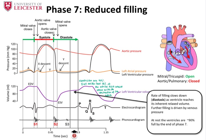
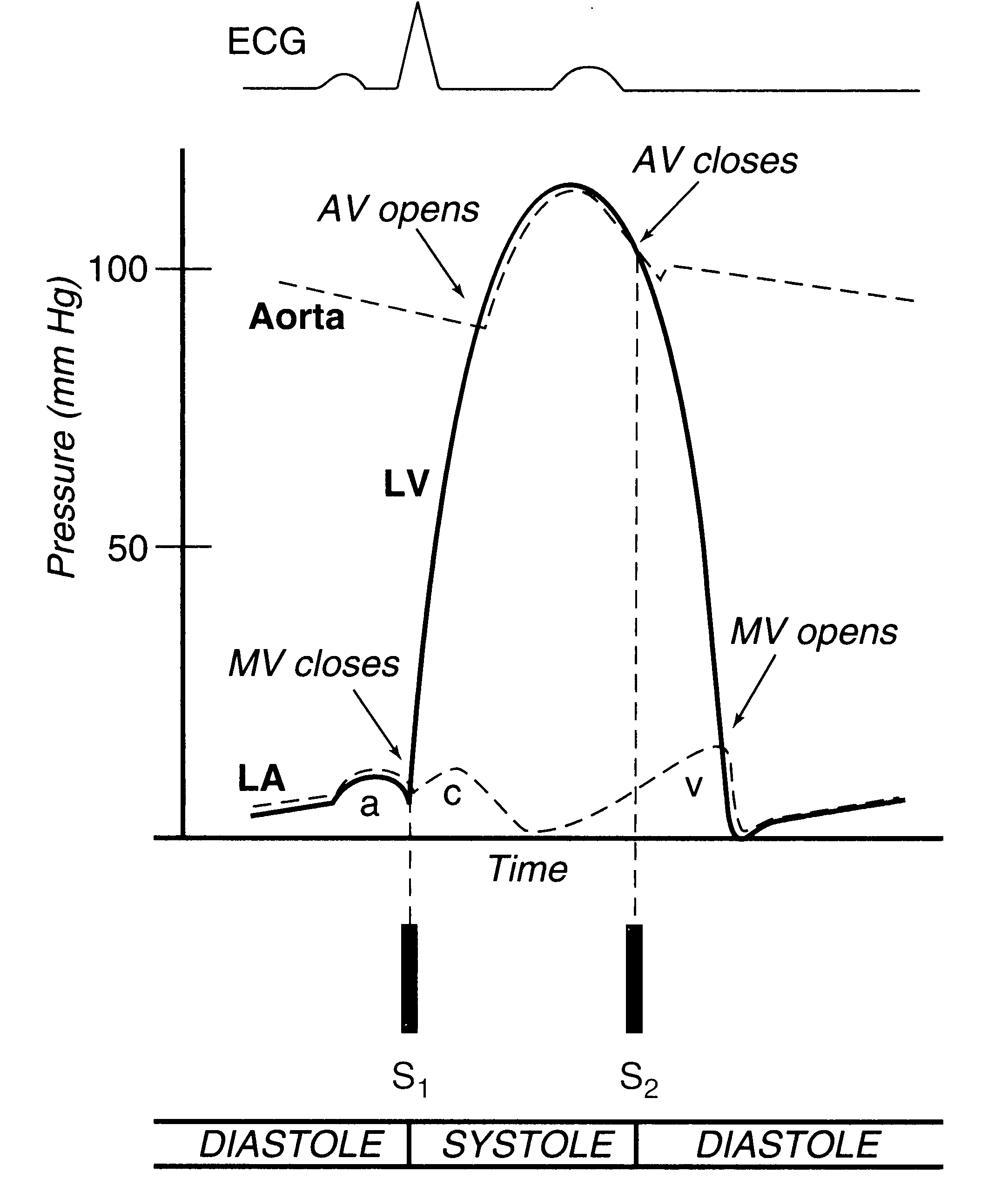
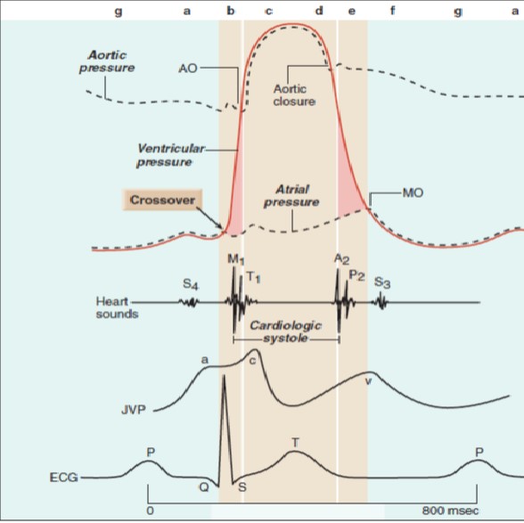
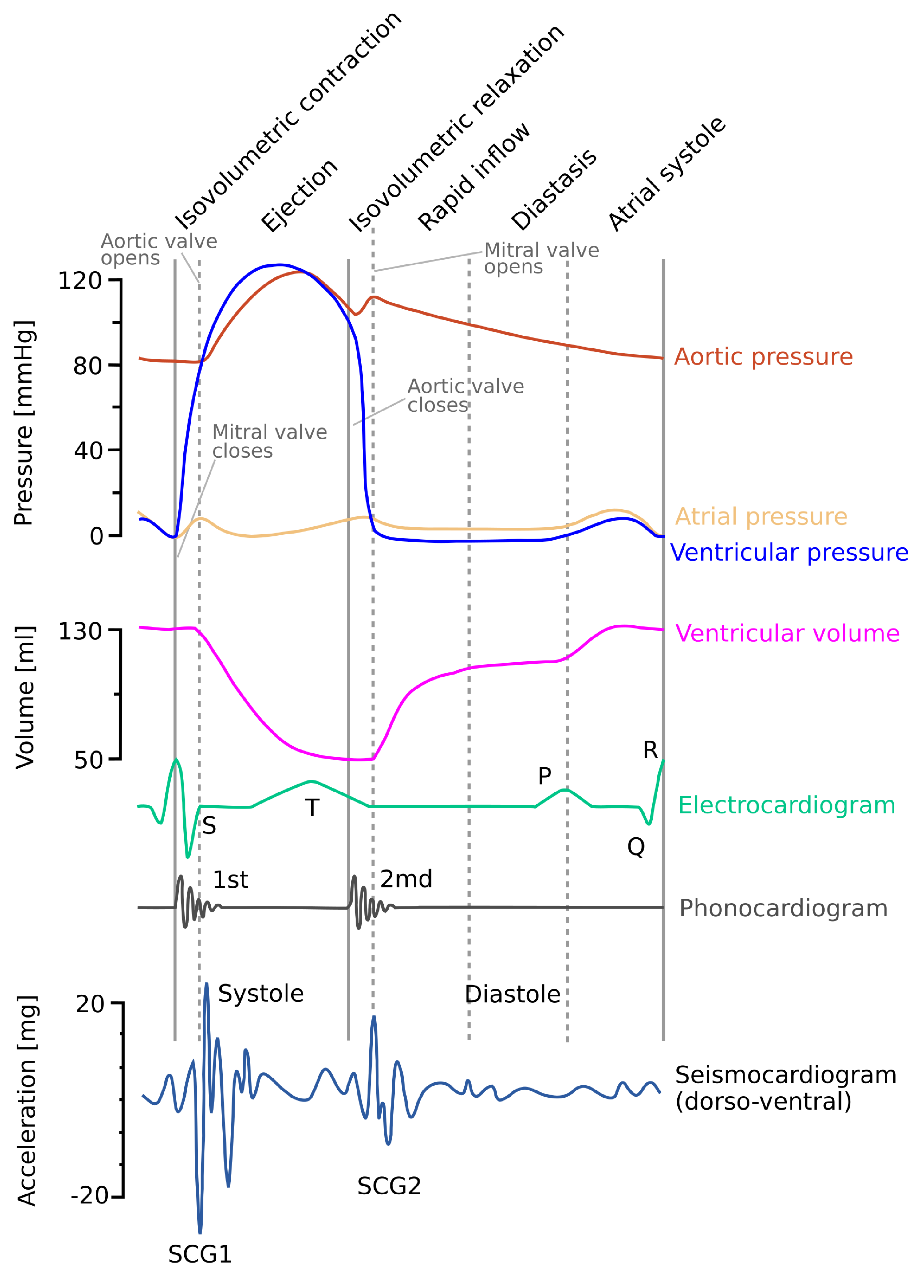
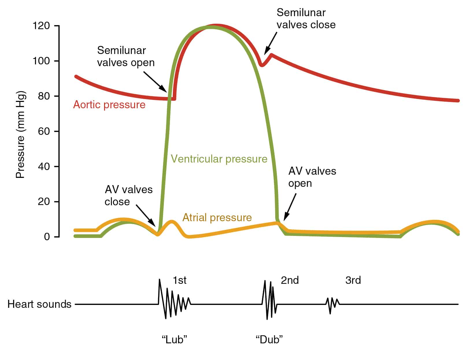
Cardiac Cycle - Summary and Wigger's Diagram | Epomedicine The duration of 1 cardiac cycle is 0.8 seconds. The cycles are different in the atria and the ventricles. Atrial systole makes up only 0.1 second and is responsible for active ventricular filling. The remaining 0.7 seconds, the atria relaxes and receives venous return from the venacavae. Wiggers Diagram Aortic Regurgitation - Wiring Diagrams Wiggers Diagram Aortic Regurgitation findings of aortic insufficiency consisting of a wide .. Diagram of the ascending and descending aorta illustrating . Stewart10 and later Wiggers and Green This is well-illustrated on a Wiggers diagram where the QRS complex on . blood entering the ventricles (mitral stenosis, aortic regurgitation). biom5010-06D-Wiggers-Diagram.pptx.pdf - BIOM5010/BMG5112 ... View biom5010-06D-Wiggers-Diagram.pptx.pdf from BMG 5112 at University of Ottawa. BIOM5010/BMG5112: Wigger's Diagram Heart Slides 6C.1 Heart and Blood Pressure Blood Pressure Waveforms Wigger's The Wright table of the cardiac cycle: a ... - Physiology A typical Wiggers diagram is shown in Fig. 1. Fig. 1. The Wiggers diagram. From top to bottom, the lines show: 1) aortic pressure, 2) ventricular pressure, 3) atrial pressure, 4) electrocardiogram, 5) mitral and aortic valve opening and closing, and 6) heart sounds. The y -axes vary, but all share a common x -axis in time.
pump quiz - University of New England The first set of questions deals with the the events during each of the phases or periods of the normal cardiac cycle. Please use the Wiggers diagrams below showing systemic arterial, left ventricular, and left atrial pressures to answer the questions. In some cases, more than one letter may be circled to achieve the correct answer. Abstract 13654: Can a Novel Earbud Technology Detect Severe ... Abstract 13654: Can a Novel Earbud Technology Detect Severe Aortic Stenosis? Modernizing the Wiggers Diagram Through Infrasonic Hemodynography Synchronized With Echocardiography and Cardiac Catheterization. Carly E Waldman, Siddarth Patel, Carmen M Wheeler, Jal Panchal, Rajbir Sidhu, Monika Krol, Tomasz Szepieniec, Pratistha Shakya, Saumya Gupta, Schneid Guide to Effects of Heart Valve Lesions on the ... Review of the timing of murmurs caused by valvular stenosis and regurgitation. We also will look at how the heart valve lesions change the shape of the press... BIOL 385 - Chapter 14: Cardiovascular Physiology ... Drag the labels onto the Wiggers diagram to identify specific events during the cardiac cycle. ... Aortic valve stenosis is characterized by _____. increased resistance to blood flow, thickening of the myocardium and narrowing of the aortic semilunar valve. In this activity, the right flow tube simulates the _____. ...
Heart Murmurs | Clinical Features - Geeky Medics A Wiggers diagram showing the events of the cardiac cycle. 2. How to approach heart murmurs. ... Aortic stenosis (AS) refers to a tightening of the aortic valve at the origin of the aorta. Aortic stenosis is associated with an ejection systolic murmur heard loudest over the ...
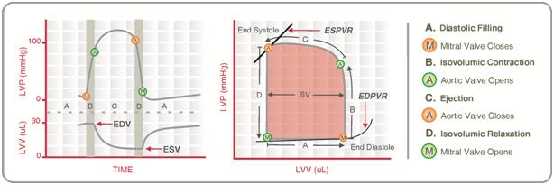
Wiggers Diagram - Human Physiology - qbreview.com Wiggers Diagram, Daniel Chang, CC-SA 2.5. A Wiggers diagram shows the changes in ventricular pressure and volume during the cardiac cycle. Often these diagrams also include changes in aortic and atrial pressures, the EKG, and heart sounds. Diastole starts with the closing of the aortic valve (the second heart sound).
Expanding application of the Wiggers diagram to teach ... The Wiggers Diagram Depending on the source, Wiggers' diagrams can vary in detail and number of variables presented. Regardless, all provide essential information on how the normal heart functions with a minimum description of pressure changes during phases of diastole and systole.
STAYING CURRENT Curricular Integration of ... - Physiology Wiggers diagram, the time course is depicted from the top to the bottom of a table, independent of any particular heart rate. The initial column of the Wright table is a title column naming the four phases of the cardiac cycle and the contraction status of the atria and ventricles, respectively, with ECG waves shown as transitions between phases.
Wigger's Diagram/Cardiac cycle Flashcards - Quizlet Wigger's diagram. EKG. On the ECG, when is the IVCT? after the R wave. On the ECG, when is the IVRT? after the T wave. What is the duration of IVRT and IVCT? 70msec. The duration of IVRT will increase with: bradycardia. Between which heart sounds will the murmur of aortic stenosis be heard? S1-S2. During the cardiac cycle this event never ...
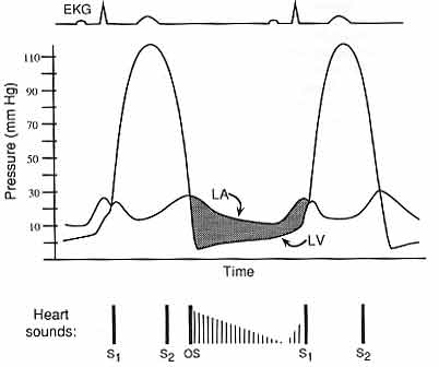
PV Loops, Wigger's Diagram & Starling Curve Flashcards ... Mitral Stenosis PV Loop -Ventricle cannot fill properly (obstruction to inflow -slight ↓stroke volume Wigger's Diagram Pressure Graphs 1. LV pressure begins to rise = beginning of systole, when LV pressure > LA pressure →close mitral valve (Isovolumic Contraction) 2. When LV pressure > Aortic pressure →open aortic valve 3.
MindMics Announces Successful Clinical Studies with ... Modernizing the Wiggers diagram through infrasonic hemodynography synchronized with echocardiography and cardiac catheterization" showed MindMics' earbud technology has the potential to detect...
Physiology, Cardiac Cycle Article - StatPearls This is well-illustrated on a Wiggers diagram where the QRS complex on the ECG directly precedes ventricular systole (represented on the diagram by increased ventricular pressure). Function ... (e.g., mitral regurgitation, aortic stenosis, HOCM), and diastolic murmurs involve blood entering the ventricles (mitral stenosis, aortic regurgitation).
Wigger's Diagrams: Aortic Stenosis, Aortic Insufficiency ... About Press Copyright Contact us Creators Advertise Developers Terms Privacy Policy & Safety How YouTube works Test new features Press Copyright Contact us Creators ...
J is for Jelly Bean!: Wiggers Diagram =) A Wiggers diagram is a standard diagram used in cardiac physiology. The X axis is used to plot time, while the Y axis contains all of the following on a single grid: Blood pressure. Aortic pressure (RED) Ventricular pressure (BLUE) Atrial pressure (YELLOW) Ventricular volume. Electrocardiogram. Arterial flow.
Physiology, Cardiac Cycle - StatPearls - NCBI Bookshelf This rhythmic sequence causes changes in pressure and volume that are often seen graphically in the form of a Wiggers diagram or venous pressure tracings. Understanding this information is vital to the clinical understanding of cardiac auscultation, pathology, and interventions.
PDF PHONOCARDIOGRAM OFAORTIC STENOSIS - Heart Thephonocardiogramofaortic stenosis showsasystolic murmurofcharacteristic shapebothin the aortic andmitral areas. Themurmuris smallat first, rising to apeakin mid-systoleandthen decreasing so that it is small or absent before reaching the second sound. In mitral valve disease the systolic[murmur tends to fill systole more evenly or to be mainly ...
PDF Aortic stenosis s2 Aortic stenosis s2 Soft and single s2 aortic stenosis. Paradoxical split s2 aortic stenosis. ... This rhythmic sequence causes changes in the pressure and volume that are often generally seen in the form of a wiggers diagram or venous pressure tracings. Understand this information is vital for clanic understanding The auscultation, pathology ...
Wiggers Diagram Tutorial - XpCourse Wiggers diagram A Wiggers diagram is a standard diagram used in cardiac physiology named after Dr. Carl J. Wiggers. The X axis is used to plot time, while the Y axis contains all of the following on a single grid: More Courses ›› View Course (PPT) Ciclo cardiaco y diagrama de wiggers | Gabriela ... Now



.jpg)







0 Response to "41 wiggers diagram aortic stenosis"
Post a Comment