42 labeled diagram of a microscope
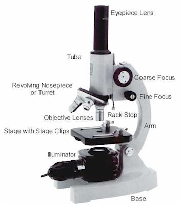
Amazing 27 things under the microscope (diagrams and descriptions) Parts of a microscope with functions and labeled diagram; Light Microscope- Definition, Principle, Types, Parts, Labeled Diagram, Magnification; Animal Cell- Definition, Organelles, Structure, Parts, Functions, Labeled Diagram, Worksheet Label Microscope Diagram Using the terms listed below, label the microscope diagram. Inventions and Inventors arm - this attaches the eyepiece and body tube to the base. base - this supports the microscope. body tube - the tube that supports the eyepiece. coarse focus adjustment - a knob that makes large adjustments to the focus.
Compound Microscope Labeled STUDY Learn Flashcards Write Spell Test PLAY Match Gravity Created by meganplocher734 Terms in this set (14) Eyepiece/Ocular lens Contains the ocular lens Body tube A hollow cylinder that holds the eyepiece. Arm Part that supports the microscope.

Labeled diagram of a microscope
A microscope is an instrument widely to magnify and resolve the image of an object that is otherwise invisible to naked eye. For resolving the details of objects, which otherwise cannot be achieved by naked eye, a microscope is used. This set of flash cards will help the student to identify the different parts and function of the microscope. Labeling the Parts of the Microscope. This activity has been designed for use in homes and schools. Each microscope layout (both blank and the version with answers) are available as PDF downloads. You can view a more in-depth review of each part of the microscope here. Jan 19, 2022 · The description given below summarize the brief description of microscope parts used to visualize the microscopic specimens such as animal cells, plant cells, microbes, bacteria, viruses, microorganisms etc. The Microscopes parts divided into three different structural parts Head, Base, and Arms. Head/Body: It contain the optical parts in the ...
Labeled diagram of a microscope. Drag and drop the pins to their correct place on the image.. Eyepiece, Light Source, Base, Stage, Stage Clips, Fine Focus, Coarse Focus, Arm, Objective Lens. Parts of a microscope with functions and labeled diagram December 2 2021 December 1 2021 by Faith Mokobi Overview of microscope Having been constructed in the 16th Century. The first page is a labelling exercise with two diagrams of the human eye. Examine the eye and identify the following parts. 28.11.2021 · Animal Cell- Definition, Organelles, Structure, Parts, Functions, Labeled Diagram, Worksheet; Amazing 27 things under the microscope (diagrams and descriptions) Types of Plant Cell- Definition, Structure, Functions, Labeled Diagram; Cell proliferation- Definition, assay, differentiation, diseases; Cell Cycle- Definition, Phases, Regulation and Checkpoints ; Plant … Draw a large diagram of an animal cell as seen through an electron microscope. Label the parts … (Myra Burns) If you want to download the image of Plant Cell Worksheet together with Structure Of Animal Cell and Plant Cell Under Microscope Diagrams, simply right click the image and choose "Save As".
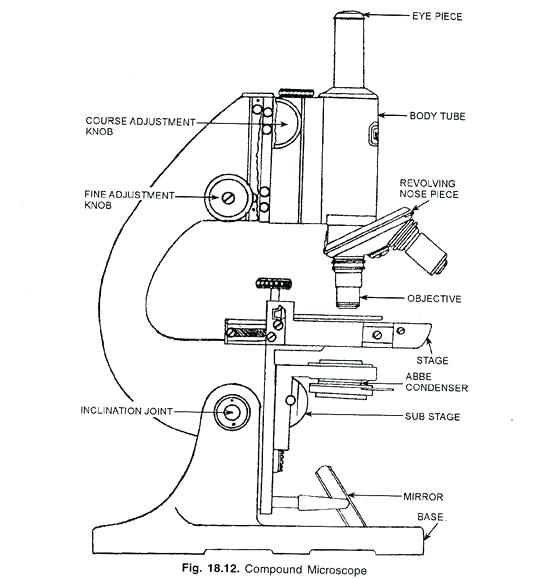
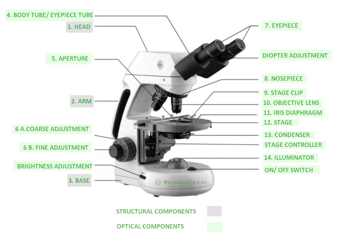
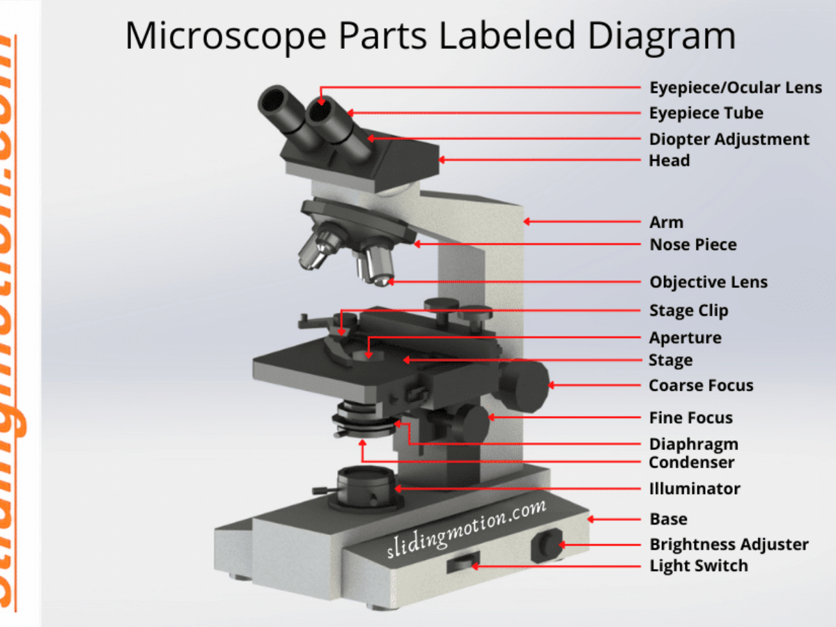
04.07.2020 · Under a light microscope Spirogyra is seen as long threadlike, green colonies called filaments that are joined end to end, without any differentiation into base and apex. Parts and their Morphology . Spirogyra. Cell Wall: Consists of three layers of which the inner two layers are made of pectin, and the outer layer is composed of cellulose. The slimy mucilaginous … Dec 24, 2021 · Figure: Diagram of parts of a microscope. There are three structural parts of the microscope i.e. head, base, and arm. Head – This is also known as the body, it carries the optical parts in the upper part of the microscope. Base – It acts as microscopes support. It also carries microscopic illuminators. labeled-diagram-of-a-microscope 1/2 Downloaded from sftp.amneal.com on January 25, 2022 by guest [MOBI] Labeled Diagram Of A Microscope Thank you definitely much for downloading labeled diagram of a microscope.Most likely you have knowledge that, people have see numerous times for their favorite books in the same way as this labeled Labeled diagram of a compound microscope Major structural parts of a compound microscope There are three major structural parts of a compound microscope. The head includes the upper part of the microscope, which houses the most critical optical components, and the eyepiece tube of the microscope.
Beside that, we also come with more related things as follows microscope parts worksheet, compound light microscope diagram labeled and compound light microscope parts worksheet. Our main objective is that these Light Microscope Diagram Worksheet images collection can be a direction for you, bring you more inspiration and most important: make ... Again, the reticulum labeled diagram shows the inner oblique pattern of the smooth muscle layer. The outer smooth muscle layer of the labeled diagram shows the cross pattern. In addition, the tunica serosa layer of the reticulum microscope labeled image shows a thin layer of loose connective tissue with numerous blood vessels. Oct 30, 2013 · Microscope labeled diagram 1. The Microscope Image courtesy of: Microscopehelp.com Basic rules to using the microscope 1. You should always carry a microscope with two hands, one on the arm and the other under the base. 2. You should always start on the lowest power objective lens and should always leave the microscope on the low power lens when you finish using it. 3. Here, I will show you all the histological structures from the cecum with a microscope slide image and labeled diagram. I will also provide the appropriate identification points for the cecum slide under the light microscope. Again, you will get a little information on the specific histological features of the cecum in a different animal.
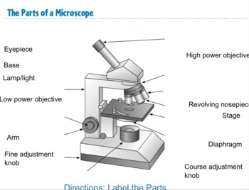
Labeled microscope worksheet answers. High power objective 6. Students label the parts of the microscope in this photo of a basic laboratory light microscope. Each microscope layout both blank and the version with answers are available as pdf downloads. When focusing a specimen you should always start with the objective.
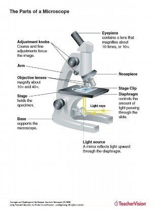
These labeled microscope diagrams and the functions of its various parts attempt to simplify the microscope for you. Drag and drop the text labels onto the microscope diagram. You can view a more in depth review of each part of the microscope here. Base this supports the microscope.
Start studying Compound Microscope Labeled. Learn vocabulary, terms, and more with flashcards, games, and other study tools.
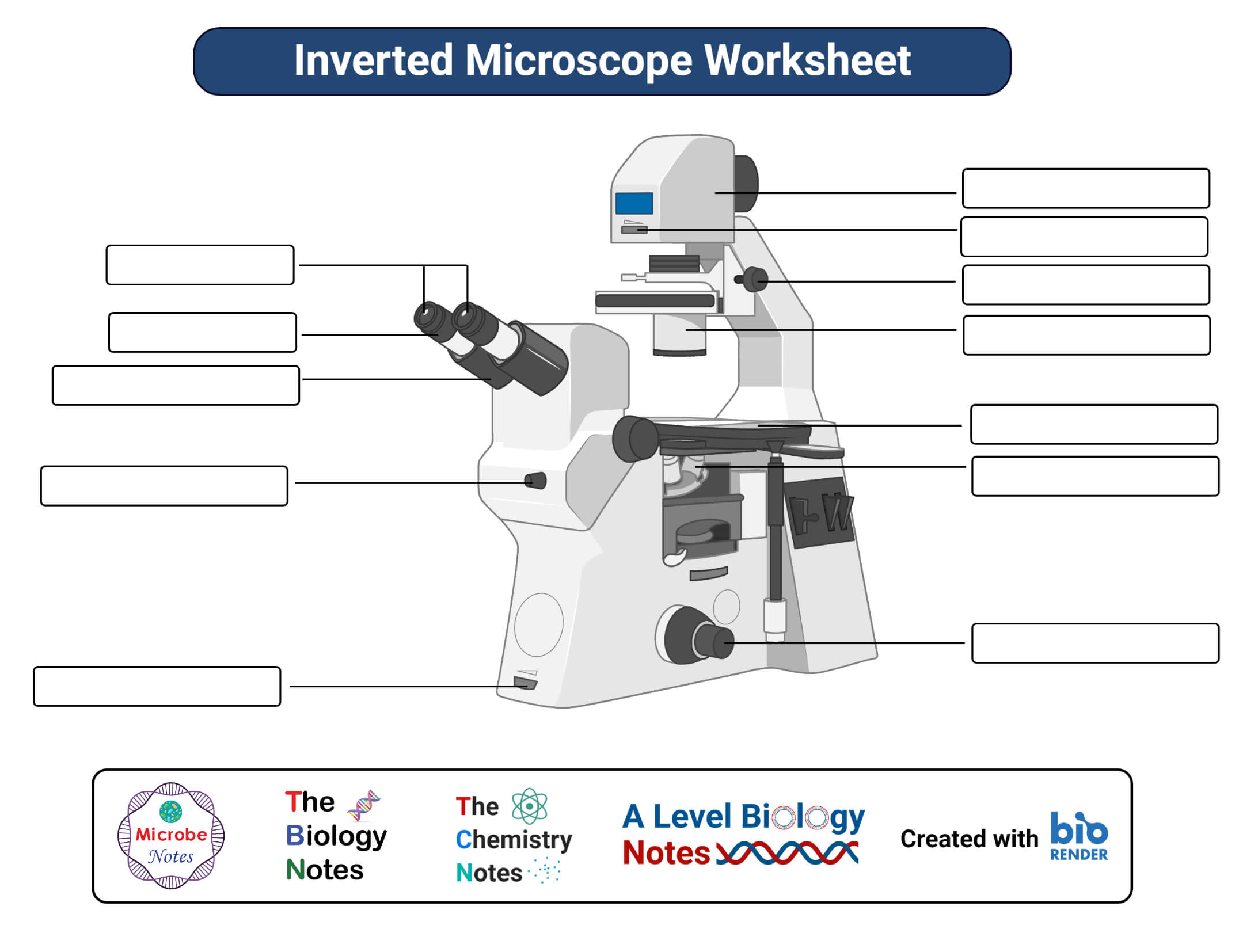
Label the parts of the microscope. You can use the word bank below to fill in the blanks or cut and paste the words at the bottom. Microscope Created by Jolanthe @ HomeschoolCreations.net eyepiece head objective lenses arm focusing knob base illuminator stage stage clips nosepiece.
labeled-diagram-of-a-microscope 1/7 Downloaded from sftp.amneal.com on January 16, 2022 by guest [Books] Labeled Diagram Of A Microscope As recognized, adventure as capably as experience practically lesson, amusement, as capably as pact can be gotten by just checking out a ebook labeled diagram of a microscope after that it is not directly done ...
Tuesday, April 20th 2021. | Diagram. Animal Cell Diagram Under Light Microscope. To make observations and draw scale. This shows a generalized animal cell under a light microscope. We all keep in mind that the human physique is amazingly elaborate and one way I discovered to comprehend it is by way of the style of human anatomy diagrams.
Jan 02, 2020 · The three basic structural components of a compound microscope are the head base and arm. These separate optical channels enable stereo or three dimensional images of the specimen....
Microscope Parts and Functions With Labeled Diagram and Functions How does a Compound Microscope Work?. Before exploring microscope parts and functions, you should probably understand that the compound light microscope is more complicated than just a microscope with more than one lens.. First, the purpose of a microscope is to magnify a small object or to magnify the fine details of a larger ...
Answer pages are only available to subscribers of EnchantedLearning.com . To subscribe to EnchantedLearning.com, click here
Those are sperm cells
So, a compound microscope with a 10x eyepiece magnification looking through the 40x objective lens has a total magnification of 400x (10 x 40). Specimen or slide: The object used to hold the specimen in place along with slide covers for viewing. ... Compound Microscope Parts, Functions, and Labeled Diagram. Parts of a Compound Microscope.
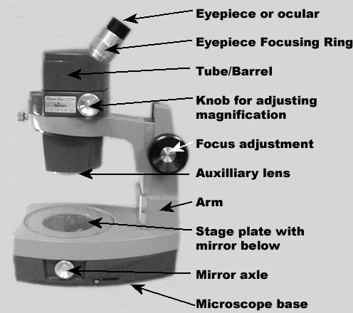
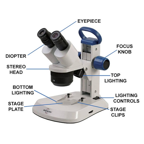
Products/Services for Labeled Diagram Of A Stereo Microscope. Microscopes - (703 companies) Microscopes are instruments that produce magnified images of small objects Microscopes are instruments that produce a magnified image of a small object. They are used in many scientific and industrial applications. Some common applications...
Labeled part diagram of a stereo microscope Major structural parts of a stereo microscope Optical components of a stereo microscope - definition and function Eyepieces Eyepiece tube Diopter adjustment ring Interpupillary Adjustment Objective Lenses Barlow lens Adjustment Knobs Light sources Stage plate Stage chips
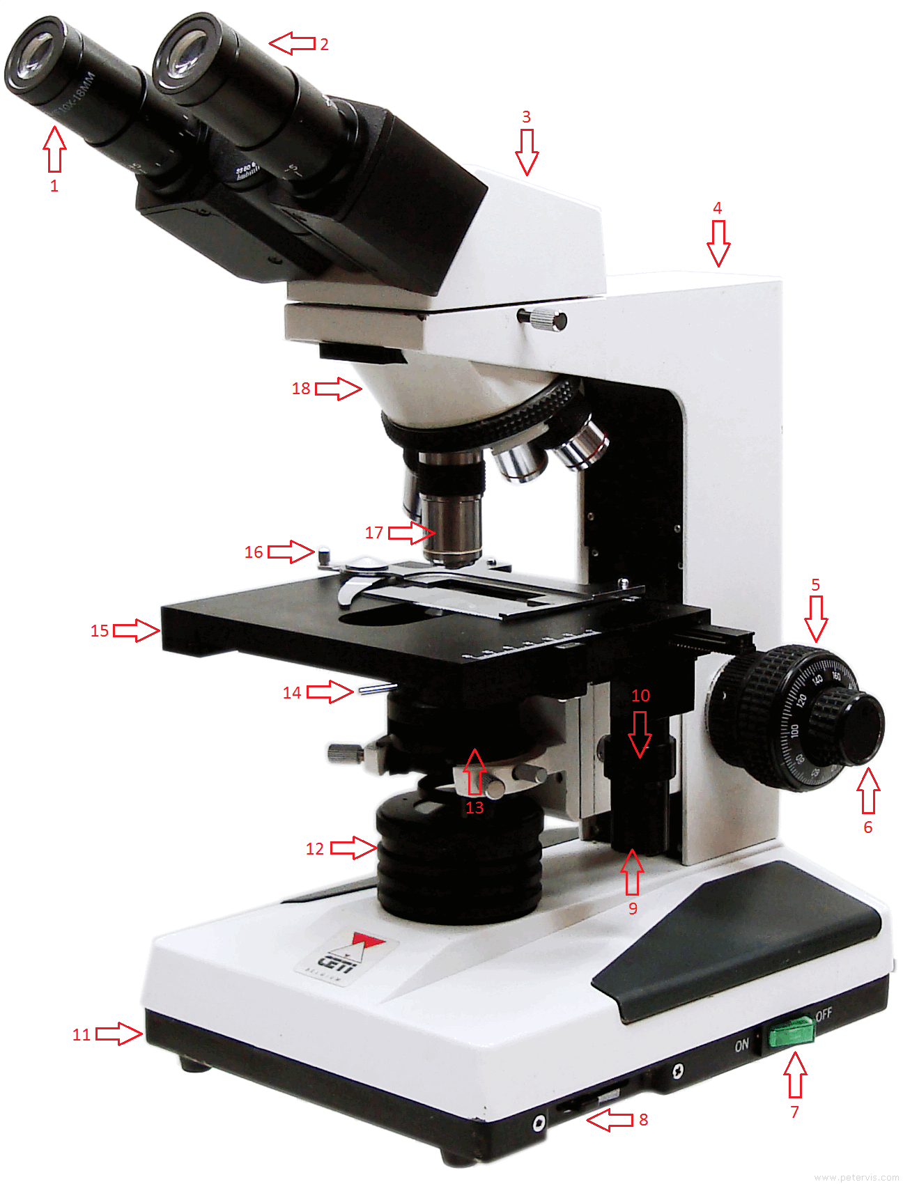
Dec 24, 2021 · To examine these small objects with high magnification microscope parts are made with special components with high accuracy. Due to that, accurate examination and results are possible to achieve. Microscope parts labeled diagram gives us all the information about its parts and their position in the microscope. Microscope Parts Labeled Diagram
Label parts of the Microscope: Answers Coarse Focus Fine Focus Eyepiece Arm Rack Stop Stage Clip www.MicroscopeWorld.com. Created Date: 20150715115425Z ...
The Parts of a Microscope (Labeled) Printable. This diagram labels and explains the function of each part of a microscope. Use this printable as a handout or transparency to help prepare students for working with laboratory equipment.
Jan 19, 2022 · The description given below summarize the brief description of microscope parts used to visualize the microscopic specimens such as animal cells, plant cells, microbes, bacteria, viruses, microorganisms etc. The Microscopes parts divided into three different structural parts Head, Base, and Arms. Head/Body: It contain the optical parts in the ...
Labeling the Parts of the Microscope. This activity has been designed for use in homes and schools. Each microscope layout (both blank and the version with answers) are available as PDF downloads. You can view a more in-depth review of each part of the microscope here.
A microscope is an instrument widely to magnify and resolve the image of an object that is otherwise invisible to naked eye. For resolving the details of objects, which otherwise cannot be achieved by naked eye, a microscope is used. This set of flash cards will help the student to identify the different parts and function of the microscope.








(159).jpg)









0 Response to "42 labeled diagram of a microscope"
Post a Comment