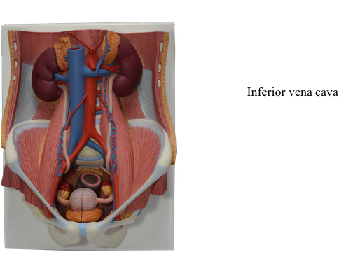
40 Inferior Vena Cava Diagram
Diagram Of Chest Area : Anatomy Of The Shoulder Muscles ... This diagram depicts the inner chest muscles in relation to the ribs and sternum . Chest area and spread to upper back. Source: legacy.owensboro.kctcs.edu. The chest, properly called the thorax, is the superior part of the trunk. Diagram of heart showing top to bottom superior vena cava, aorta, . quizlet.com › 283874553 › inferior-vena-cava-diagramInferior Vena Cava Diagram - Quizlet Start studying Inferior Vena Cava. Learn vocabulary, terms, and more with flashcards, games, and other study tools.
In the given diagram which one is vena cava ? In the given diagram which one is vena cava ? Books. Physics. NCERT DC Pandey Sunil Batra HC Verma Pradeep Errorless. Chemistry. NCERT P Bahadur IIT-JEE Previous Year Narendra Awasthi MS Chauhan. Biology. NCERT NCERT Exemplar NCERT Fingertips Errorless Vol-1 Errorless Vol-2. Maths. NCERT RD Sharma ...
Inferior vena cava diagram
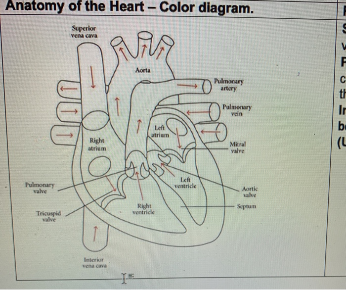
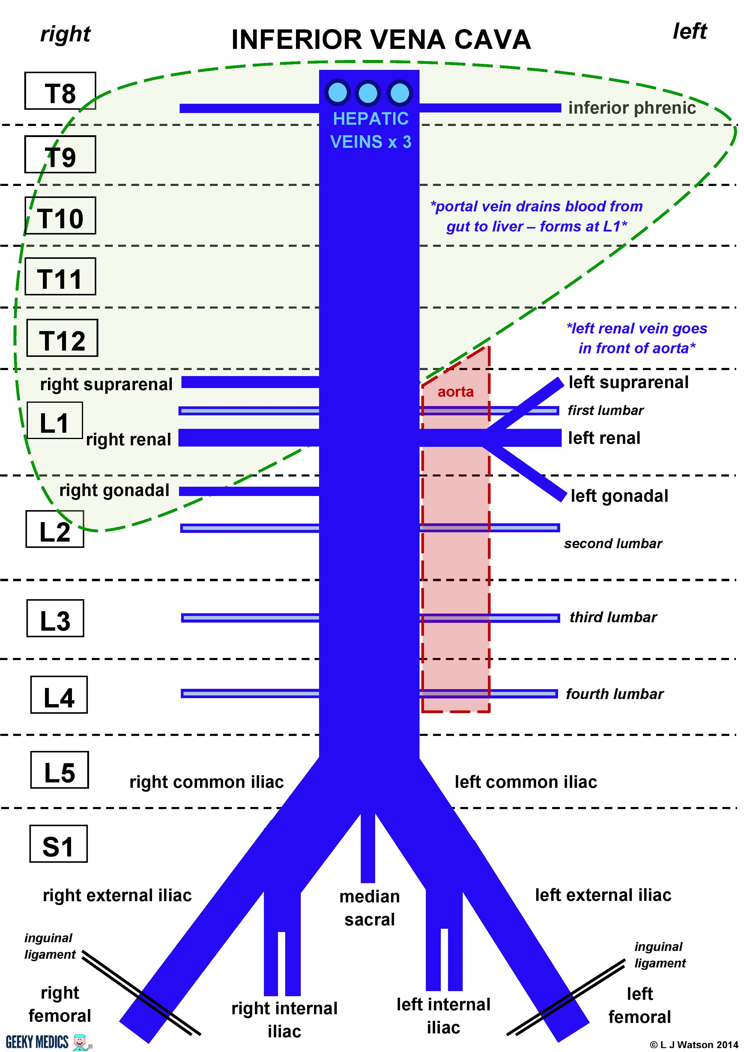
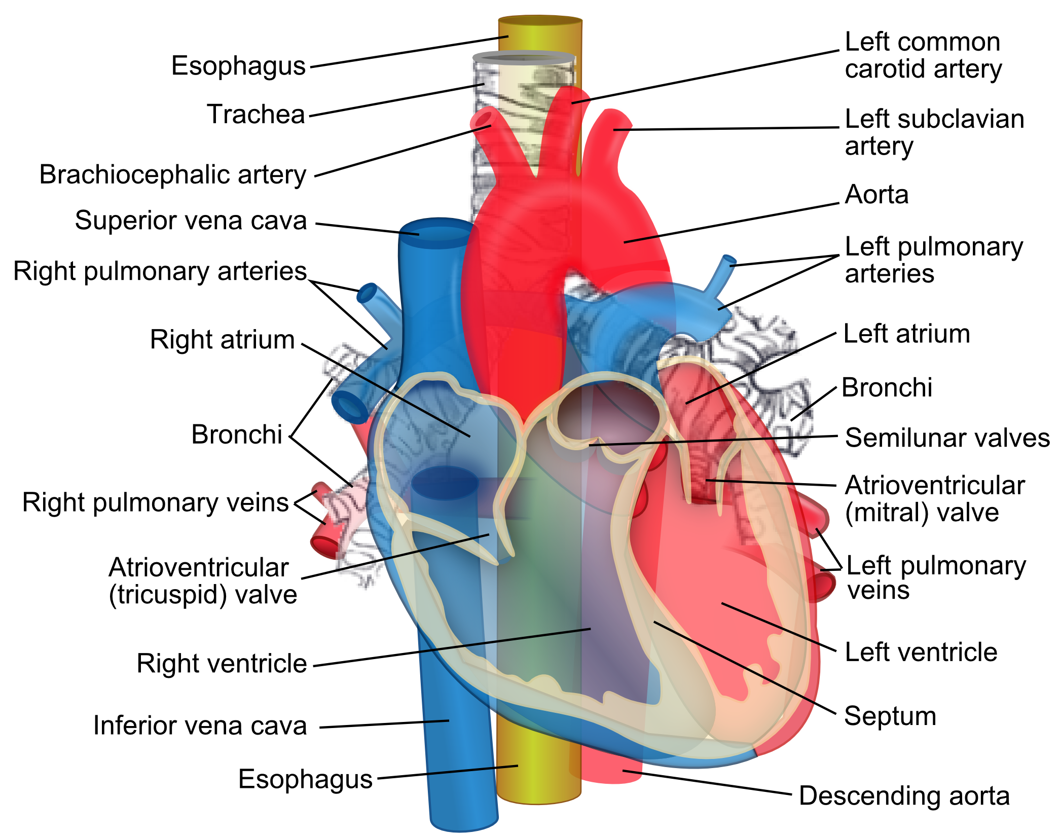
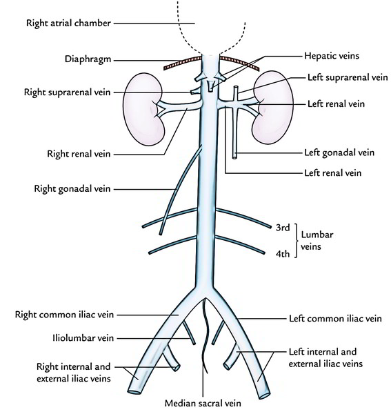
Quiz 9: Circulatory System Anatomy and Basic Functions ... Label the Heart Diagram using the word list provided. Each answer is worth 1/2 mark each. Word List: Superior vena cava Right pulmonary artery Right pulmonary veins. Right atrium Rt. AV Valve (Tricuspid) Right ventricle. Inferior vena cava Left pulmonary artery Left pulmonary veins. Inferior vena cava: Anatomy and function - Kenhub The inferior vena cava (IVC) is the largest vein of the human body. It is located at the posterior abdominal wall on the right side of the aorta. The IVC's function is to carry the venous blood from the lower limbs and abdominopelvic region to the heart.. The inferior vena cava anatomy is essential due to the vein's great drainage area, which also makes it a hot topic for anatomy exams. › inferior-vena-cava-diagramInferior Vena Cava Diagram - Anatomy Note Jul 11, 2019 · Inferior Vena Cava Diagram. In this image, you will find hepatic veins, inferior phrenic vein, portal vein, left renal vein, left suprarenal vein, left gonadal vein, right gonadal vein, right renal vein in it. You may also find right suprarenal vein, aorta, left common iliac vein, right common iliac vein, left external iliac vein, median sacral ...
Inferior vena cava diagram. Diagram of Human Heart and Blood Circulation in It | New ... The aorta is about an inch wide and supplies oxygen-rich blood to the body's cells. The used blood them moves back and is collected into the two veins, the superior vena cava that receives blood from your upper body, and the inferior vena cava that receives blood from your lower body. scienceforyou.netlify.app › inferior-vena-cava-diagramInferior vena cava diagram | scienceforyou Inferior vena cava diagram. Inferior Vena Cava Diagram.There are several key points to take away from this diagram. Although the vena cava is very large in diameter its walls are incredibly thin due to the low pressure exerted by venous blood. › image_dige07 › card26Inferior Vena Cava - Anatomy Pictures and Information Dec 15, 2016 · Inferior Vena Cava. The inferior vena cava is the largest vein in the human body. It collects blood from veins serving the tissues inferior to the heart and returns this blood to the right atrium of the heart. Although the vena cava is very large in diameter, its walls are incredibly thin due to the low pressure exerted by venous blood. What is the Celiac Artery? (with pictures) The celiac artery provides the spleen and pancreas, as well as other organs, with blood. Most arteries carry oxygenated blood. The deoxygenated blood from the celiac artery returns to the heart and lungs via the inferior vena cava. The celiac artery supplies blood and nutrients to the spleen, among several other organs.
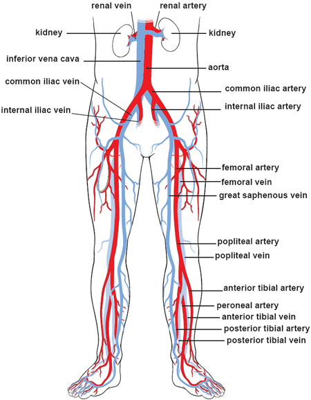
Inferior Vena Cava Anatomy Branches Function Human Anatomy ... Inferior vena cava. the inferior vena cava (ivc) is the largest vein of the human body. it is located at the posterior abdominal wall on the right side of the aorta. the ivc's function is to carry the venous blood from the lower limbs and abdominopelvic region to the heart . the inferior vena cava anatomy is essential due to the vein's. Anatomy, Thorax, Superior Vena Cava - StatPearls - NCBI ... The superior vena cava is a large, significant vein responsible for returning deoxygenated blood collected from the body back into the heart. It is present within the superior and middle mediastinum. The superior vena cava handles the venous return of blood from structures located superior to the diaphragm. The inferior vena cava handles venous return from the portion of the body inferior to ... Blood vessels of abdomen and pelvis : Anatomy ... - Kenhub The inferior vena cava is the headmaster of the veins department. It collects all the blood from the abdomen, pelvis and lower limbs and carries it to the right atrium of the heart.. The IVC is formed by merging of the left and right common iliac veins at the L5 vertebral level, just in front of the aortic bifurcation.. The inferior vena cava then ascends to the right of the abdominal aorta ... Fetal Circulation Diagram | Fetal Blood Flow & Circulatory ... The inferior vena cava takes blood to the right atrium of the heart Through a series of shunts and openings, the blood flows through the heart bypassing the lungs.
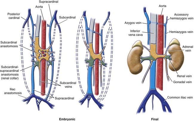
Duplication of inferior vena cava | Radiology Reference ... The inferior vena cava has a convoluted development during the 7-10 th weeks of gestation 4. posterior cardinal vein appears first but forms only the distal IVC i.e. iliac bifurcation. subcardinal veins (2) appear next, left subcardinal vein regresses, and right subcardinal vein forms the suprarenal IVC. Inferior Vena Cava (IVC) - Geeky Medics Overview of the inferior vena cava. The IVC is formed by the union of the right and left common iliac veins.It conveys systemic venous blood from the lower limbs and pelvis, the undersurface of the diaphragm and parts of the abdominal wall.The IVC does not drain blood from the gut.. Course of the IVC. The IVC begins in the abdomen at L5 and ends in the thorax at T8, where it enters the ... Diagram of capillaries veins and arteries - Summarized by ... The superior vena cava brings blood from the head and arms to the heart, and the inferior vena cava brings blood from the abdomen and legs into the heart. Capillaries are narrow-diameter tubes that can accommodate red blood cells in single-file lines and are the points for the exchange of nutrients, waste, and oxygen with tissues at the ... Human Body Urinary System Diagram - Studying Diagrams These biochemical reactions taking place in the body may produce toxic wastes which. Because the inferior vena cava is on. Organisms carry out biochemical reactions to produce energy. Human PhysiologyThe Urinary System 4 Renal Vein The renal veins are veins that drain the kidneyThey connect the kidney to the inferior vena cava.
Solved - -- - MITU T yumuy co, WILllli, allu Culinny ruil ...
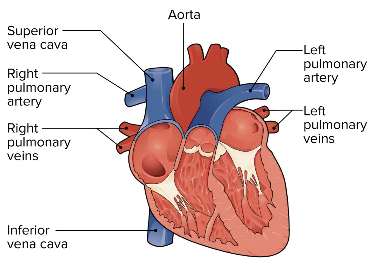
CBSE class 10th science sample paper - satnaz.com 1- pulmonary artery - Carries blood to the body. 2- Superior and inferior vena cava - carries blood to lungs. 3- Aorta - Carries blood from lungs. 4- Pulmonary veins - Carries blood from lungs. A person was found to have reduced level of reabsorption of water and glucose due to kidney damage.

Congenital absence of inferior vena cava. | Semantic Scholar
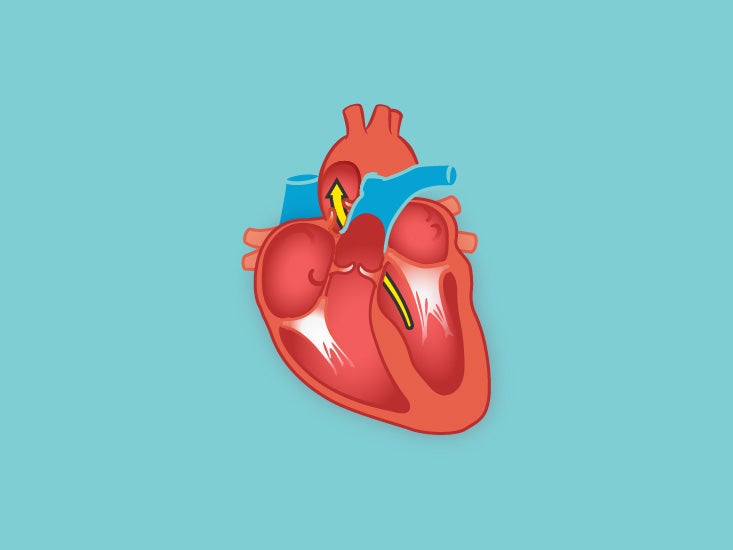
Flow Of Deoxygenated Blood Through The Heart - Etfatehran.net The pathway of blood flow through the heart begins as blood comes from the body and enters the heart through the superior and inferior vena cava indicated by the yellow star in the diagram below. Superior and inferior vena cavae and the coronary sinus 2. The right atrium receives deoxygenated blood through the superior and inferior vena cavas ...
Inferior Vena Cava (IVC) | Geeky Medics
Heart Anatomy: Labeled Diagram, Structures, Blood Flow ... Superior/Inferior Vena Cava. Now that we understand the blood flow to and from the heart, we can discuss the final structures. The first 2 structures are responsible for carrying deoxygenated blood from the body to the right side of the heart (right atrium). They are known as the superior vena cava and inferior vena cava.

Superior and inferior vena cava Diagram | Quizlet
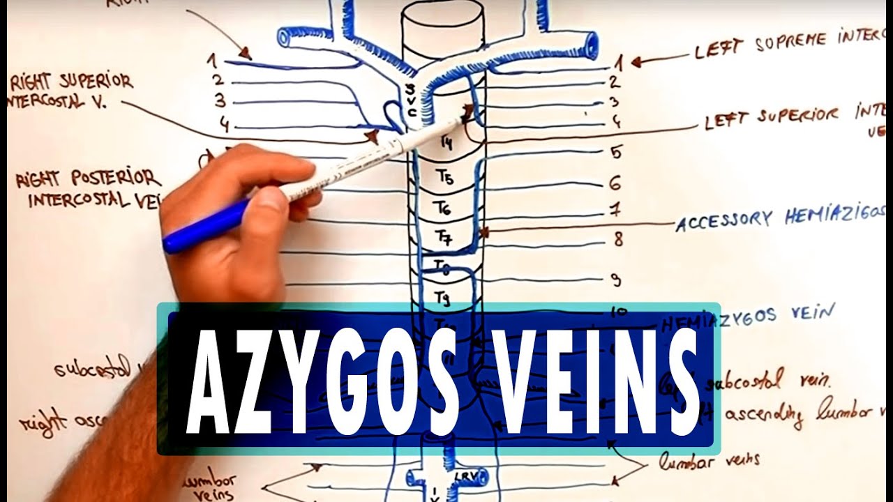
which blood vessel does the azygos vein empty into ... Most of the time, the azygos vein originates from a portion of the inferior vena cava that is posterior to the renal veins. It ascends through the posterior mediastinum to the level of T4 before arching upward over the right pulmonary hilum, as shown in the diagram.

Inferior Vena Cava (IVC) | Geeky Medics
Inferior vena cava | Radiology Reference Article ... Gross anatomy. The inferior vena cava is formed by the confluence of the two common iliac veins at the L5 vertebral level. The IVC has a retroperitoneal course within the abdominal cavity.It runs along the right side of the vertebral column with the aorta lying laterally on the left. Various other veins drain into the IVC along its course before it passes through the diaphragm at the caval ...
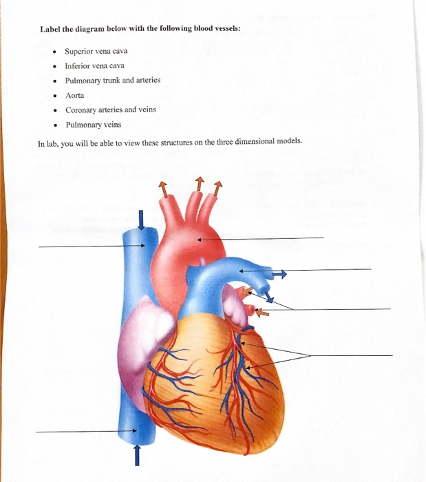
Solved Label the diagram below with the following blood ...
A Labelled Diagram Of The Human Body - Studying Diagrams Know the structure of the heart Labelled diagram. The right atrium receives deoxygenated blood from upper part of the body by superior vena cava and inferior vena cava collects blood from he lower part of the body. The organ where protein digestion starts.
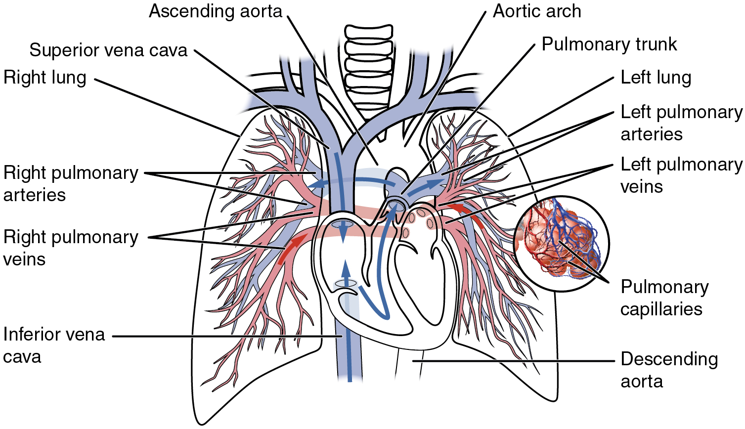
Circulatory Pathways – Anatomy and Physiology
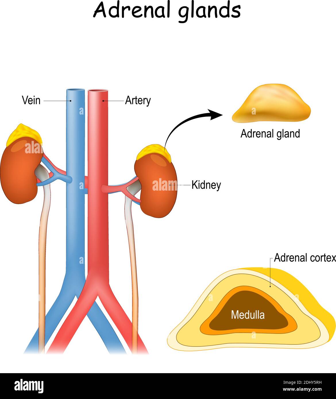
Lymphatic Drainage of the Liver and ... - Barnard Health Care The deep networks along the hepatic veins drain into the lymph nodes around the inferior vena cava (IVC) above the diaphragmatic opening through which the IVC passes (Fig. 6-5). This nodal group is known as the IVC terminal nodes or juxtaphrenic nodes. Fig. 6—2. Deep pathways of lymphatic drainage of the liver.

inferior vena cava | anatomy | Britannica
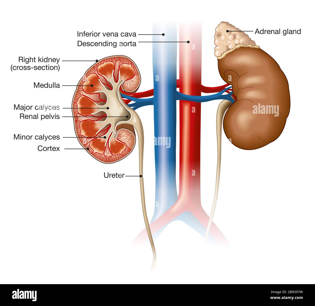
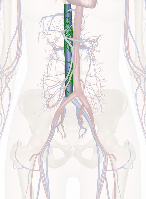
Venous Drainage of the Abdomen - TeachMeAnatomy The inferior vena cava is the common convergence of venous drainage from all structures below the diaphragm. It is located on the posterior abdominal wall; anteriorly to the vertebral column and to the right of the abdominal aorta. The vessel is formed by the union of the common iliac veins at the L5 vertebral level.

Tributaries of the inferior vena cava and lumbar veins [4 ...
diagram of how blood flows through the heart - Lisbdnet.com 38 Box Diagram, Labels of Heart, and Blood Flow through Heart; ... What is the order of blood flow through the heart assume we are starting at the superior and inferior vena cava? The blood enters the heart from the body through the superior vena cava and the inferior vena cava. Then the blood enters the right atrium chamber of the heart.
Venae cavae - Wikipedia
Heart Blood Flow | Simple Anatomy Diagram, Cardiac ... Diagram: Blood flow through the right side of the heart involving the following cardiac structures: superior vena cava (SVC), inferior vena cava (IVC), right atrium (RA), tricuspid valve (TV), right ventricle (RV), pulmonary valve (PV), and main pulmonary artery (PA).
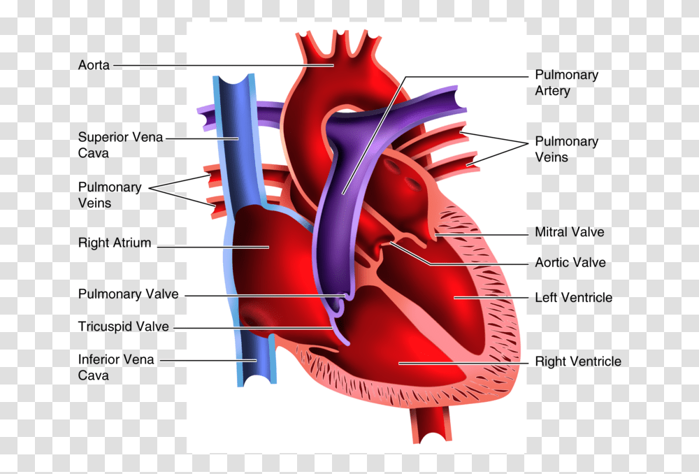
Diagram Of The Human Heart Bs Inferior Vena Cava, Plot ...
What is Portal Circulation? (with picture) - Info Bloom The hepatic veins leave the liver, emptying into a vein called the inferior vena cava, which is part of the systemic blood circulation that carries blood back to the heart. From the inferior vena cava, blood is delivered to the right upper chamber of the heart, or right atrium, before being ejected from the right lower chamber, or ventricle ...
Aorta And Vena Cava High Resolution Stock Photography and ...
Inferior Vena Cava Syndrome - StatPearls - NCBI Bookshelf Inferior vena cava syndrome (IVCS) is a sequence of signs and symptoms that refers to obstruction or compression of the inferior vena cava (IVC). The pathophysiology of IVCS is similar to superior vena cava syndrome (SVCS) because of the presence of an underlying process that inhibits venous return to the right atrium. IVCS is not a primary diagnosis because it is often caused by other ...

Circulatory System: Blood Flow Pathway Through the Heart ...
Frontiers | PUTH Grading System for Urinary Tumor With ... Introduction. Renal tumors have the tendency to involve the venous system ().In some patients the thrombus can invade the renal vein as well as the inferior vena cava (IVC) and even involve the right atrium (RA) (2-4).It was reported that 4% to 10% of the patients with renal cell carcinoma (RCC) had venous thrombus and 1% RCC patients had thrombus involving the RA (5, 6).

69 Inferior Vena Cava Stock Photos, Pictures & Royalty-Free ...
Physiology, Pulmonary Circulation - PubMed Pulmonary circulation is the system of transportation that shunts de-oxygenated blood from the heart to the lungs to be re-saturated with oxygen before being dispersed into the systemic circulation. Deoxygenated blood from the lower half of the body enters the heart from the inferior vena cava while …

Inferior Vena Cava Clip Art - Royalty Free - GoGraph
› inferior-vena-cava-diagramInferior Vena Cava Diagram - Anatomy Note Jul 11, 2019 · Inferior Vena Cava Diagram. In this image, you will find hepatic veins, inferior phrenic vein, portal vein, left renal vein, left suprarenal vein, left gonadal vein, right gonadal vein, right renal vein in it. You may also find right suprarenal vein, aorta, left common iliac vein, right common iliac vein, left external iliac vein, median sacral ...

Inferior Vena Cava Tributaries: (A) Geeky Medics ...
Inferior vena cava: Anatomy and function - Kenhub The inferior vena cava (IVC) is the largest vein of the human body. It is located at the posterior abdominal wall on the right side of the aorta. The IVC's function is to carry the venous blood from the lower limbs and abdominopelvic region to the heart.. The inferior vena cava anatomy is essential due to the vein's great drainage area, which also makes it a hot topic for anatomy exams.
Abdominal Aorta and the Inferior Vena Cava | Radiology Key
Quiz 9: Circulatory System Anatomy and Basic Functions ... Label the Heart Diagram using the word list provided. Each answer is worth 1/2 mark each. Word List: Superior vena cava Right pulmonary artery Right pulmonary veins. Right atrium Rt. AV Valve (Tricuspid) Right ventricle. Inferior vena cava Left pulmonary artery Left pulmonary veins.

PC182. National Trends in Inferior Vena Cava Filter Placement ...
Illustrations of the Blood Vessels
:max_bytes(150000):strip_icc()/heart_and_major_vessels-5820b6ba3df78cc2e887becd.jpg)
Superior and Inferior Venae Cavae
Inferior Vena Cava (IVC) – Earth's Lab
Skill Lab Learning ::
Mediastinum and Great Vessels | Concise Medical Knowledge

Vein Systemic venous system Circulatory system Anatomy ...

Figure: Inferior Vena Cava Filters: One Way to Prevent ...
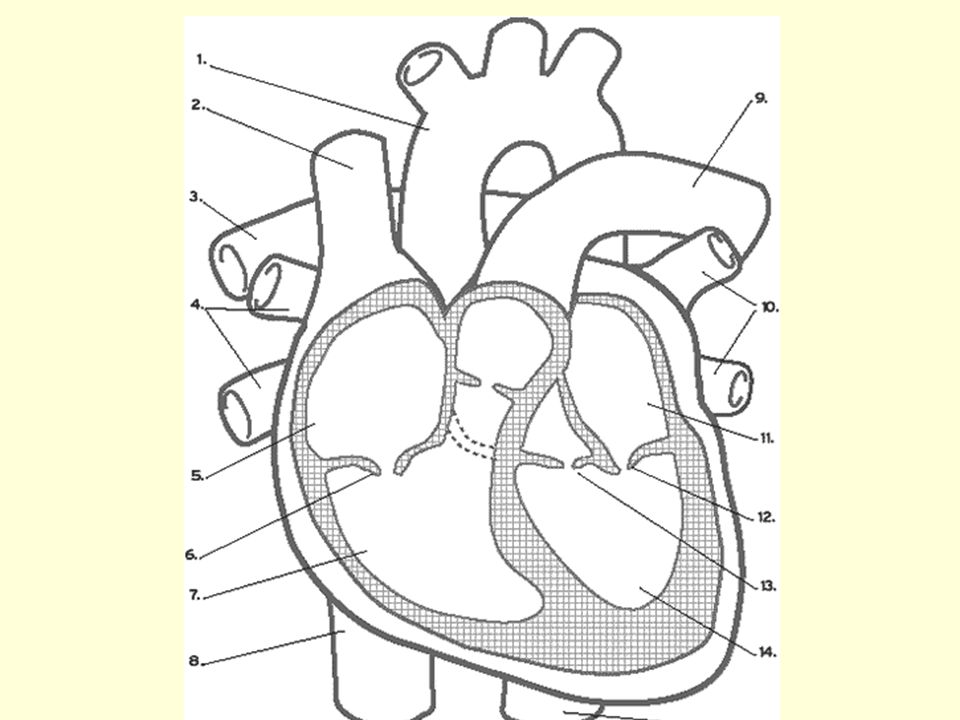
Diagram Word Bank Superior Vena Cava Inferior Vena Cava Right ...

Cava Inferior Vena Stock Illustrations – 74 Cava Inferior ...

Anatomy of a Human Heart

JaypeeDigital | eBook Reader

Inferior Vena Cava Diagram | Quizlet
Inferior Vena Cava Function, Anatomy & Definition | Body Maps
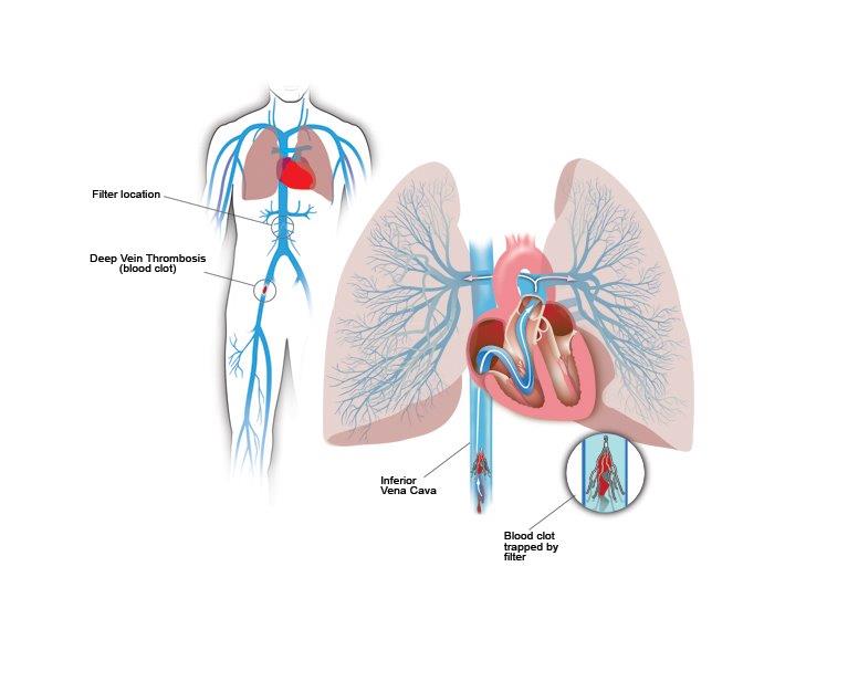
Inferior Vena Cava Filters | Center for Vein Care
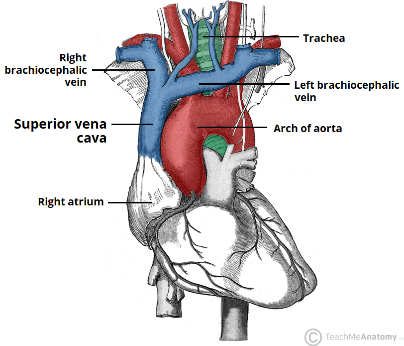
The Superior Vena Cava - TeachMeAnatomy

Portal vein Hepatic veins Hepatic portal system Inferior vena ...
Inferior Vena Cava and its tributaries - Anatomy Tutorial ...

The tributaries of the superior and inferior vena cava ...

Schematic drawing of embryologic development of the inferior ...
Solved] 9. Using the correct terminology from the list below ...
Inferior Vena Cava High Resolution Stock Photography and ...
Inferior Vena Cava - Anatomy Pictures and Information
0 Response to "40 Inferior Vena Cava Diagram"
Post a Comment